
Start here - basic information
Newsletter
BioVis NewsLetter September 2026
ELECTRON MICROSCOPY
The BioVis EM node has been doing its part in the creation of this new book ” Om Krokodiler - Deras hörsel och andra sinnen - Vad kan vi lära av dem?” written by Kalle Melkersson, Hao Li and Helge Rask-Andersen. Check in some cool electron microscope images of crocodile auditory hair cells!
Interested in doing SEM, STEM or elemental mapping (EDS) - Please contact us for further information. We are happy to assist with the biological sample preparation!
FLOW CYTOMETRY
Flow Cytometry Applications & Analysis (Course)
Robin Lindsay, researcher at the Department of Medical Cell Biology, is delivering this highly engaging and insightful course on flow cytometry (October).
This course introduces the fundamentals of flow cytometry and shows how it can be used in research. It combines basic concepts with practical work, so participants both learn the theory and try it out themselves.
By the end of the course, students will be able to plan and carry out different types of flow cytometry experiments relevant to their own projects. They will design antibody panels tailored to their experimental needs and gain confidence working with data—from handling raw output to creating clear, publication-ready figures.
Throughout the course, participants will practice designing experiments and analysing flow cytometry data, helping them build skills they can apply directly in their research.
To apply follow the link
SPECTRAL Flow Cytometry
The Beckman Coulter Cytoflex MOSAIC is installed, manuals are in the making to ensure a smooth introduction in October. Together with Beckman Coulter we are currently planning a Kick-off-event in October.
Flow Educations sources
For those interested to learn about the different aspects about FlowCytometry the internet is your helper: Check out the education sources: Open Flow Cytometry and those Post-it.
LIGHT MICROSCOPY
Stellaris is Live
Stellaris, now with Live Cell imaging capabilities: as of June 2026, BioVis offers two new stage top incubation chambers that are specially designed for imaging. A smaller chamber for looking at chamber slide or small petri dishes and a larger chamber for multi-well plates. The Ibidi Stage Top Incubators – Silver Line – allow for the regulation and recording of temperature, gas concentrations and humidity. BioVis is also acquiring a tank of compressed N2 gas for running experiments under hypoxic conditions. Currently, the system is set up for use on the Rudbeck Stellaris confocal microscope. Reach out - ross.smith@igp.uu.se – to learn more and for help getting started.
Acknowledgements
Acknowledgements
Customers of BioVis are required to acknowledge BioVis, and relevant BioVis staff where applicable, in all publications, presentations, and other public disclosures that include data generated through or supported by BioVis services.
Example: The BioVis platform of Uppsala University was used to conduct experiments using “technique /Instrument”, supported by “Staff name “.
General Information
- General Information
BioVis offers a wide range of different instruments. In general BioVis has three nodes: Electron Microscopy, Flow Cytometry (and Cell Sorting), Light Microscopy. More information is available under the Technology section.
We are mainly situated at Rudbeck Lab and have a small site at BMC. The BMC site offers one Flow Cytometer and one Confocal Microscope at B11, third floor. UU Campus card is needed to access the room. You reach the room following this mazemap link.
An Introduction to the Instruments given by BioVis Staff is priority for all customers, before using BioVis´Instruments independently.
Full Service can be booked, where BioVis staff is handling the customers´ processed sample on the Instrument.
An Access card is needed to enter BioVis Premises.
Approved users have access to the facility 24 hours a day 7 days a week. However after 17:00, and during weekends, access is restricted to experienced users who are familiar with the instruments and can preform some trouble shooting themselves.
BioVis may be closed for all customer during shorter periods, concerning vacation. This will always be announced at least two months in advance giving you plenty of time to plan your experiments accordingly.
Report all instrument problems directly to BioVis staff and make a note in the logbook every time you have used an instrument.
Bring the consumables you will need to perform your analysis, like cell-strainers, tubes, glass slides and cover slips etc. They are not included in the fee.
Access Card
- Entrance card & Access
In order to enter BioVis premises at Rudbeck Lab, you need to have a valid access card. Customers of IGP and Rudbeck need only access granted on their existing cards. An access card can be obtained via BioVis. The access card is personal and must not be lent to a colleague. Access cards are given out to customers who have received Instrument introductions and use our instruments.
Please contact ross.smith@igp.uu.se for more details regarding access cards. Observe that your cards will be valid for one year in total. Have the access card form xlsx, 13 kB. ready to be sent to Ross.
Since 240411 we have a FlowCytometer at the BMC, B11 third floor. See General Information menue for more details.
Instrument Introductions
- Instrument Introductions
BioVis gives introductions to its instruments once per month. The introduction contain in-depth lectures and hands-on sessions on the instrument. Introduction to the instrument is a priority to be an independent customer. You can apply for introductions under Introductions.
iLab Booking System
BioVis is using iLab as its current Booking system.
Each customer to BioVis services (for Light Microscopy and Flow Cytometry) needs to have a valid account.
Make sure
- your Research group PI has a valid account including invoice details
- you are accepted to use your PIs invoice details and funding
The following documents give you more detailed information:
- Document for Research group PI pdf, 114 kB.
- Document for Researcher (customer BioVis) pdf, 139 kB.
- Document for external customers (not UU) pdf, 132 kB.
Additional help
More detailed instructions can be found by clicking on the “HELP” link in the upper right hand corner or by navigating to our iLab Help Site. For any questions not addressed in the Helpsite, click on the “HELP” link in the upper right hand corner and submit a ticket, or email itsupport@uu.se or ilab-support@agilent.com.
For bookings in iLab use the link iLab bookings at BioVis
Service & Prices
Instruments
BioVis offers a wide range of different instruments. In general BioVis has three nodes: Electron Microscopy, Flow Cytometry (and Cell Sorting), Light Microscopy. More information is available under the Technology section.
Services
- Flow Cytometry, Cell Sorting, Light Microscopy
BioVis offers its customers independ use of instruments or Full Service. For independent use of Instruments, customers are obliged to have been trained by BioVis staff. Apply for it under Instrument Introductions. Full Service can be booked. Here staff is handling the instrument, using your (processed) sample. BioVis offers also Consulting to discuss and guide projects concerning experimental design (in relation to instrument use).
- Electron Microscopy
BioVis offers its customer a full service mode from sample preparation to imaging.
Prices for Services
BioVis is mainly financed by the MedFarm Faculty of Uppsala University and service prices are subsidized for UU customers. A detailed price list can be seen below BioVis has a three level price setting: Uppsala University, Swedish Academia, Full cost price. Within this levels you will find Independent use and Full service, as well as different categories like Courses etc.
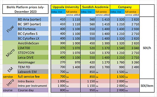
Current BioVis Platform Prices (Adopted July 2023)
Prices are shown as SEK/hr or SEK/course
If BioVis staff accept a full service request, an additional fee is added (shown in right column)
Current BioVis Platform Prices (Adopted July 2023)
| BioVis Node | Equipment | Uppsala University | Swedish Academia | Other | |||
|---|---|---|---|---|---|---|---|
| Independent Use | Full Service | Independent Use | Full Service | Independent Use | Full Service | ||
| Flow | +700 | +850 | +1500 | ||||
| BD Aria (sorter) | 410 | 1110 | 560 | 1410 | 1320 | 2820 | |
| BC SRT (sorter) | 410 | 1110 | 560 | 1410 | 1210 | 2710 | |
| BC Cytoflex S | 400 | 1100 | 550 | 1400 | 1430 | 2710 | |
| BC Cytoflex LX | 400 | 1100 | 550 | 1400 | 1320 | 2820 | |
| Microscopy | |||||||
| AxioScan Z.1 (slidescanner) | 300 | 1000 | 450 | 1300 | 1320 | 2820 | |
| LSM700 (confocal) | 370 | 1070 | 520 | 1370 | 1540 | 3040 | |
| Stellaris (confocal) | 370 | 1070 | 520 | 1370 | 1540 | 3040 | |
| STEDYCON (super-resolution) | 370 | 1070 | 520 | 1370 | 1210 | 2710 | |
| Leica DIVE (multiphoton) | 400 | 1100 | 550 | 1400 | 1210 | 2710 | |
| AxioImager (widefield, upright) | 270 | 970 | 420 | 1270 | 760 | 2260 | |
| Electron Microscopy | |||||||
| TEM Hitachi | 700 | 1400 | 850 | 1700 | 990 | 2490 | |
| Labwork EM | 700 | N/A | 850 | N/A | 1500 | N/A | |
| Analysis | |||||||
| IMARIS (3D-4D analysis software) | 100 | 200 | 1000 | ||||
| Huygens (deconvolution) | 70 | 100 | 1000 | ||||
| FlowJo (workstation) | |||||||
| Courses | |||||||
| Intro Basics | 1000 | N/A | 1150 | N/A | 2000 | N/A | |
| Instrument Intro (per instrument) | 1000 | N/A | 1150 | N/A | 2000 | N/A | |
Data Safety / Biological Safety
- Biosafety
All work involving biosafety class 2 organisms on BioVis instruments must be performed according to Uppsala University regulations. You are obliged to inform us about any potential hazards.
The use of lentiviral vectors has been increasing because they allow highly efficient gene delivery in a wide variety of cell types. Samples of lentivirus-transduced cells pose a higher degree of hazard than other samples submitted for analysis. Have such samples in culture for one week so virus activity is ceased.
- Data storage
We cannot be held responsible for the misuse, loss and manipulation of data stored on our computers. You are allowed to store data temporarily on our computers. Always transfer your data to a trusted storage medium. Details on how to handle your data will be found on the instrument computer desktop. We export all stored data from instrument computers on a regular basis. Our data share computer is a public computer, all BioVis users will have access to your data there. We reserve the right to delete older data folders without prior notice if storage media owned by BioVis reach their capacity limits.
PhD & Customer Courses
BioVis offers also courses in Image Analysis based on the usage of Image J. BioVis runs two PhD courses, Methods for Cell Analysis , and Introduction to Image Analysis Software.
You find more information under Courses.