
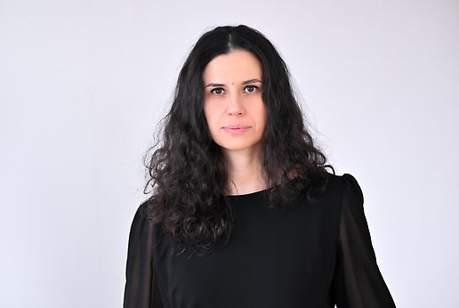
Sanja Vickovic
- E-post:
- sanja.vickovic@igp.uu.se
- Besöksadress:
- BMC, Husargatan 3
751 22 Uppsala - Postadress:
- IGP / BMC
Box 815
751 08 Uppsala
Kort presentation
Mitt laboratorium utvecklar innovativa teknologier och beräkningsmässiga ramverk för att spatialt kartlägga cellulär funktion i komplexa vävnader, med särskilt fokus på cancer, åldrande och neurodegenerativa sjukdomar. Vi utvecklar spatiala multiomik-plattformar och analysmetoder som integrerar transkriptomiska, epigenetiska och proteomiska mätningar med hög upplösning, vilket möjliggör en systematisk kartläggning av vävnadsarkitektur och genreglering in situ. Tillämpade på patienthärledda och e
Nyckelord
- Spatial Transcriptomics
- Digital Pathology
- Genomics
- AI/ML
Biografi
10/2021, Broad Institute Wallenberg Fellow, Computational Biology, Broad Institute of MIT and Harvard, Cambridge
02/2017, PhD, Genetic Engineering, Royal Institute of Technology
07/2011, MS, Molecular Biomedicine, Royal Institute of Technology
07/2009, BS, Bioengineering, University of Zagreb, Zagreb
Forskning
cSplotch: Convolutional hierarchical Bayes for modelling tissue structures
“cSplotch,” is a novel algorithm that combines cellular and spatial data to track gene expression patterns linked to aging and tissue structure. With cSplotch, we tackle age-related gene expression decline by constructing an atlas of cellular changes across the colon from birth through old age, using both single-nuclear RNA sequencing and spatial transcriptomics. To integrate these data and address the shortcomings of each modality, “cSplotch”, a Bayesian statistical model of cellular gene expression is conditioned on important sample covariates such as age, tissue region, and sex. cSplotch leverages histological image features to share information across disparate tissues and data modalities, and allows for testing of hypotheses of differential expression across age, spatial location, or other sample covariates. Through this approach, we have identified key genes and cellular functions that deteriorate with age, highlighting potential targets for therapeutic interventions aimed at preserving colon function in older populations. cSplotch is now available to the research community as a tool for similar studies in other tissues.
PySeq 2500: An open source toolkit for repurposing HiSeq 2500 sequencing systems as versatile fluidics and imaging platforms
PySeq2500 is an open source Python code base and flow cell design which enables the conversion of the Illumina HiSeq 2500 instrument into an open platform suitable for programmable applications. Adaptable PySeq2500 protocols allow for experimental designs involving simultaneous 4-channel image acquisition, temperature control, reagent exchange, stable positioning, and preservation of sample integrity over extended periods of experimentation. As a demonstration of the attainable level of automation in complex, multi-day workflows, we have utilized the PySeq2500 system for the unattended execution of iterative indirect immunofluorescence and immunoSABER imaging. In essence, our automated software control approach utilizes readily available antibodies to produce highly multiplexed maps of cell types and pathological features in tissue sections through multiple cycles of staining and imaging. Hence, PySeq2500 serves to facilitate the development and implementation of cutting-edge fluidics-linked imaging methods on a commonly accessible benchtop system for non-specialists.
Hudson: an ecosystem for high-throughput optical mapping using decomissed sequencers
We have expanded upon our previous efforts in repurposing decommissioned Illumina instrumentation. Our new package, known as Hudson, stands as a comprehensive spatial-omics ecosystem available to researchers and laboratories alike. This ecosystem incorporates all necessary steps, ranging from flow cell generation to tissue handling for multiplexed antibody staining and detection, followed by repeated rounds of imaging, raw image processing, cell segmentation, cell type calling, and spatial neighborhood analysis. In order to tackle the issue of spatial niche discovery in spatial proteomics data, we are introducing a novel model known as single-cell embedded latent Dirichlet allocation (sceLDA). This addition to the Hudson ecosystem combines the scalability of deep generative models with the interpretability of topic modeling, effectively categorizing imaged cells into anatomical regions and determining cell type mixtures associated with various disease states. Similar to other workflows for image processing and analysis, our pipelines are entirely free, open source, and modular tools designed to process and analyze multiple types of imaging data.
SHM-seq: spatial host-microbiome sequencing analysis package
Mucosal and barrier tissues, such as the gastrointestinal tract, respiratory system, or integumentary system, are composed of a complex interplay between cells and microbes that create a tight ecosystem. This highly specialized niche effectively prevents pathogen colonization and fosters host-microbiome symbiosis. Therefore, it is imperative to comprehensively study these networks at a molecular and cellular level in order to gain a deeper understanding of homeostasis and disease processes. To that end, we present spatial host-microbiome sequencing (SHM-seq): a cutting-edge, all-sequencing based technique that enables the simultaneous capture of tissue histology, polyadenylated RNAs, and bacterial 16S sequences using spatially barcoded glass surfaces. We have successfully applied this approach to a mouse model of the gut, implementing a deep learning algorithm for data mapping and revealing distinct spatial niches defined by cellular composition and bacterial geography. Our findings demonstrate that subpopulations of gut cells exhibit unique gene programs in different microenvironments, which are highly influenced by the presence of regional commensal bacteria and significantly impact host-bacteria interactions. As such, the utilization of SHM-seq holds great promise for furthering our understanding of native host-microbe interactions in both healthy and pathological states.
INLAomics: Spatial generalized linear mixed models (GMMM) methods for multiomic analysis using Integrated Nested Laplace Approximations (INLA):
Integrating spatial transcriptomics with antibody-based proteomics allows for the comprehensive investigation of biological regulation within intact tissue architecture. Nevertheless, current approaches for spatial multi-omics integration often rely on dimensionality reduction or autoencoders, both of which overlook spatial context and limit interpretability. Additionally, these methods face challenges with scalability. In response to these limitations, we have developed INLAomics, a multivariate hierarchical Bayesian framework that accurately models protein abundance in tissue sections by incorporating histological features and latent spatial factors inferred from spatial transcriptomics data. Furthermore, INLAomics supports two key applications: (1) identifying spatial gene co-expression programs to construct interpretable gene-protein networks and (2) predicting spatial protein expression in tissues without proteomics measurements. Through its application on diverse datasets, INLAomics uncovers previously unidentified gene-protein associations and exhibits significant enhancements in protein prediction accuracy compared to models that consider each modality independently. This framework is not only computationally efficient but also biologically interpretable, providing a scalable solution for the integrative analysis of extensive spatial multi-omics data.
Celery is a comprehensive tool that aims to streamline and enhance the process of annotating spatial transcriptomic data in a web-based platform. This platform offers the unique capability of allowing multiple users to collaboratively annotate or view the same sample, with all progress automatically synced with a centralized database. Not only does this facilitate efficient teamwork, but it also ensures the accuracy and consistency of annotations across projects. Celery currently supports data from Visium and Pyseq 4i, with partial support for original ST. The platform is divided into several tabs to cater to various annotation needs. For instance, the "Project Summaries" tab provides a quick overview of the annotation status for each tissue in the assigned projects, while the "Curated Annotations" tab allows users to manually perform tissue annotations using defined anatomical regions. The application also offers the option to import annotations from Loupe Browser in .csv format and export the current annotations as a .csv file. Additionally, the "Computed Annotations" tab, although still a work in progress, offers predictions made by ML models, which can be copied over to the "Curated Annotations" tab. The platform also facilitates gene expression analysis, allowing users to select specific genes or calculated QC metrics from Visium-style data and view them simultaneously. Furthermore, registered users can conveniently upload projects and samples to the database using the command line tool provided, with the only prerequisite being a user token that can be obtained by clicking the "Get Token" button. Overall, Celery offers a user-friendly and efficient solution for annotating spatial transcriptomic data, making it a valuable resource for researchers and scientists working in this field.
Tiger: In silico tissue generation and power analysis for spatial omics
As spatially resolved multiplex profiling of RNA and proteins becomes more common, it is important to understand the available statistical power for testing specific hypotheses during experiment planning and analysis. In an ideal scenario, a predictive model would determine the sample size needed for a wide range of spatial experiments. However, uncertainty about the number of significant spatial features and the complex nature of spatial data analysis make this task challenging. In this work, we identify several key parameters that should be considered when designing a spatial omics study with adequate statistical power. We introduce a method for generating customizable in silico tissues (ISTs) and demonstrate how to use these simulations alongside spatial profiling datasets to build an exploratory computational framework for spatial power analysis. Our results show that this framework is versatile, working effectively with different types of spatial data and tissue samples. Although our focus is on spatial power analysis, these simulated tissues also hold promise for benchmarking and optimizing other spatial analysis methods.
3dst: Three-dimensional spatial transcriptomics uncovers cell type dynamics in the human rheumatoid arthritis synovium
This repository contains all code related to 3D spatial transcriptomics analysis in the rheumatoid arthritis synovium. Inflamed joints in rheumatic conditions are complex tissues, where multiple cell types are dynamically recruited and interact in various ways within confined areas. Traditionally, the synovium in rheumatoid arthritis has been studied using immunostaining methods or by examining homogenized tissue for molecular profiles. In our approach, Spatial Transcriptomics is used, which involves labeling tissue-resident RNA in situ with barcodes across the entire transcriptome. This method allows us to examine local tissue interactions at the site of chronic synovial inflammation. We present detailed spatial RNA-Seq data that reveals cell type-specific patterns surrounding organized clusters of infiltrating leukocytes. By combining morphological characteristics with high-throughput spatial transcriptomics, our workflow aims to improve statistical power and provide better insights for monitoring disease severity and treatment responses in both seropositive and seronegative rheumatoid arthritis.
Hdst: High-definition spatial transcriptomics for in situ tissue profiling
This public repository contains all code associated with High-definition Spatial Transcriptomics (HDST). Tissue function relies on both spatial and molecular features, but few methods can capture these details at high resolution. Our HDST technique addresses this by using a dense array of spatially barcoded beads to collect RNA directly from tissue sections. Each experiment yields several hundred thousand RNA-linked spatial barcodes at a 2-μm resolution, as demonstrated in mouse brain and primary breast cancer samples. This approach enables a detailed, high-resolution spatial analysis of cells and tissues.
Sm-omics: SM-Omics: An automated platform for high-throughput spatial multi-omics
The spatial organization of cells and molecules plays a key role in tissue function in homeostasis and disease. Spatial Transcriptomics (ST) has recently emerged as a key technique to capture and positionally barcode RNAs directly in tissues. Here, we advance the application of ST at scale, by presenting Spatial Multiomics (SM-Omics) as a fully automated high-throughput platform for combined and spatially resolved transcriptomics and antibody-based proteomics.
Media
Karolinska Institutet. (2016, July 1). New method provides better information on gene expression [News release]. Retrieved from https://news.ki.se/new-method-provides-better-information-on-gene-expression
RNA-Seq Blog. (2016, October 14). MASC-seq – Massive and parallel expression profiling using microarrayed single-cell sequencing [News article]. Retrieved from https://www.rna-seqblog.com/masc-seq-massive-and-parallel-expression-profiling-using-microarrayed-single-cell-sequencing
Oncology Central. (2016, October 20). Novel microarray‐based method for examining single leukemia cells could improve treatment [Press release]. Retrieved from https://www.oncology-central.com/novel-microarray-based-method-examining-single-leukemia-cells-improve-treatment
News Medical. (2019, October 1). New method for studying individual cells could lead to earlier detection of diseases [News release]. News Medical. Retrieved from https://www.news-medical.net/news/20191001/New-method-for-studying-individual-cells-could-lead-to-earlier-detection-of-diseases.aspx
Technology Networks. (2019, April 5). ALS progression revealed by high-res spinal cord study [News]. Technology Networks. Retrieved from https://www.technologynetworks.com/neuroscience/news/als-progression-revealed-by-high-res-spinal-cord-study-317741
Simons Foundation. (2019, April 4). How ALS progresses on genetic and cellular level revealed by high-res spinal cord study [Press release]. Simons Foundation. Retrieved from https://www.eurekalert.org/news-releases/596133
Science Daily. (2019, April 4). How ALS progresses on genetic and cellular level revealed by high-res spinal cord study [News]. ScienceDaily. Retrieved from https://www.sciencedaily.com/releases/2019/04/190404143659.htm
New York Genome Center. (2019, April 4). Researchers create multidimensional gene expression atlas, offering unprecedented detail into ALS disease progression [News release]. New York Genome Center. Retrieved from https://www.nygenome.org/news-events/news/researchers-create-multidimensional-gene-expression-atlas-offering-unprecedented-detail-intoals-disease-progression/
Devitt, J. (2019, April 4). Researchers create multidimensional gene expression atlas, offering unprecedented detail into ALS disease progression [News release]. New York University. Retrieved from https://www.nyu.edu/about/news-publications/news/2019/april/researchers-create-multidimensional-gene-expression-atlas--offer.html
Heger, M. (2019, March 4). Spatial genomics a hot topic at AGBT as 10x Genomics, NanoString prepare to launch products [News]. GenomeWeb. Retrieved from https://www.genomeweb.com/sequencing/spatial-genomics-hot-topic-agbt-10x-genomics-nanostring-prepare-launch-products#.ZEpRZexBxrw
KTH Royal Institute of Technology. (2019, October 1). High-resolution RNA-sequencing enables detection of disease at its earliest stages [News article]. Retrieved from https://phys.org/news/2019-10-high-resolution-rna-sequencing-enables-disease-earliest.html
NanoString Technologies, Inc. (2019, February 25). NanoString to host inaugural Spatial Genomics Summit on Wednesday, February 27th at the 2019 Advances in Genome Biology and Technology (AGBT) Conference [Press release]. Retrieved from https://www.globenewswire.com/news-release/2019/02/25/1741353/26898/en/NanoString-to-Host-Inaugural-Spatial-Genomics-Summit-on-Wednesday-February-27th-at-the-2019-Advances-in-Genome-Biology-and-Technology-AGBT-Conference.html
RNA-Seq Blog. (2019, October 9). High-resolution RNA-sequencing enables detection of disease at its earliest stages [Commentary]. Retrieved from https://www.rna-seqblog.com/high-resolution-rna-sequencing-enables-detection-of-disease-at-its-earliest-stages/
Morton, C. (2021, February 16). Swedes garner Method of Year by showing both gene expression and location of single cells [News article]. Retrieved from https://nshg-pm.org/swedes-garner-method-year-showing-both-gene-expression-and-location-single-cells
Finall, A. (2022, December). 3rd edition advances in single-cell and spatial analysis [Technical report]. Retrieved from https://www.researchgate.net/publication/366438036_3RD_EDITION_ADVANCES_IN_SINGLE-CELL_AND_SPATIAL_ANALYSIS
Favrot, L. (2022, July 1). Champion hockey player’s road to science [News article]. Columbia Biomedical Engineering. Retrieved from https://www.bme.columbia.edu/champion-hockey-players-road-science
Knut and Alice Wallenberg Foundation. (n.d.). New technology to understand cancer development in the intestine [Press release]. Retrieved from https://www.wallenberg.org/
Tay, A. (2021, March 31). Spatial transcriptomics is transforming immuno-oncology research [News article]. Retrieved from https://www.labmanager.com/insights/spatial-transcriptomics-is-transforming-immuno-oncology-research-25470
Columbia University. (2023). Sanja Vicković awarded a two‐year grant from Target ALS [Press release]. Retrieved from https://cancerdynamics.columbia.edu/news/sanja-vickovic-awarded-two-year-grant-target-als
Knut and Alice Wallenberg Foundation. (2023). New technique set to improve understanding of colorectal cancer [Press release]. Retrieved from https://kaw.wallenberg.org/en/research/new-technique-set-improve-understanding-colorectal-cancer
Yanai, I. (2023, July 19). What do you most hope spatial molecular profiling will help us understand? Part 2. Cell Systems, 14. https://doi.org/10.1016/j.cels.2023.05.009
Columbia University. (2025). Vicković lab publishes new study on colon aging [Press release]. Retrieved from https://cancerdynamics.columbia.edu/news/vickovic-lab-publishes-new-study-colon-aging-nature-biotechnology
Uppsala University. (2025). Unreaveling the complexities of colon aging [Press release]. Retrieved from https://www.uu.se/en/department/immunology-genetics-and-pathology/research/research-news/archive/2025-10-27-unravelling-the-complexities-of-colon-aging
Press related to outreach
New York Genome Center. (2023, May 8). NYGC women reflect on women’s history month 2023 [News release]. Retrieved from https://www.nygenome.org/nygc-women-reflect-on-womens-history-month-2023/
Favrot, L. (2023, August 30). The Herbert and Florence Irving Institute for Cancer Dynamics at Columbia University Celebrates Five Years [News release]. Retrieved from https://cancerdynamics.columbia.edu/news/herbert-and-florence-irving-institute-cancer-dynamics-columbia-university-celebrates-five
Favrot, L. (2023, March 1). The IICD Celebrates Women's History Month: Spotlight IICD Scientists [News article]. Retrieved from https://cancerdynamics.columbia.edu/news/womens-history-month-spotlight-iicd-scientists
Francisco, M. (2023, July 14). Five questions with Sanja Vicković. Nature Biotechnology, 41, 1031. https://doi.org/10.1038/s41587-023-01856-y
Publikationer
Senaste publikationer
-
From slices to deep dishes: spatial transcriptomics and translatomics of thick tissue blocks
Ingår i Nature Methods, s. 2500-2502, 2025
-
Tissue and cellular spatiotemporal dynamics in colon aging
Ingår i Nature Biotechnology, 2025
- DOI för Tissue and cellular spatiotemporal dynamics in colon aging
- Ladda ner fulltext (pdf) av Tissue and cellular spatiotemporal dynamics in colon aging

-
SenNet recommendations for detecting senescent cells in different tissues
Ingår i Nature reviews. Molecular cell biology, s. 1001-1023, 2024
-
Spatial host-microbiome sequencing reveals niches in the mouse gut
Ingår i Nature Biotechnology, s. 1394-1403, 2024
- DOI för Spatial host-microbiome sequencing reveals niches in the mouse gut
- Ladda ner fulltext (pdf) av Spatial host-microbiome sequencing reveals niches in the mouse gut
-
In silico tissue generation and power analysis for spatial omics
Ingår i Nature Methods, s. 424-+, 2023
- DOI för In silico tissue generation and power analysis for spatial omics
- Ladda ner fulltext (pdf) av In silico tissue generation and power analysis for spatial omics
Alla publikationer
Artiklar i tidskrift
-
From slices to deep dishes: spatial transcriptomics and translatomics of thick tissue blocks
Ingår i Nature Methods, s. 2500-2502, 2025
-
Tissue and cellular spatiotemporal dynamics in colon aging
Ingår i Nature Biotechnology, 2025
- DOI för Tissue and cellular spatiotemporal dynamics in colon aging
- Ladda ner fulltext (pdf) av Tissue and cellular spatiotemporal dynamics in colon aging
-
Spatial host-microbiome sequencing reveals niches in the mouse gut
Ingår i Nature Biotechnology, s. 1394-1403, 2024
- DOI för Spatial host-microbiome sequencing reveals niches in the mouse gut
- Ladda ner fulltext (pdf) av Spatial host-microbiome sequencing reveals niches in the mouse gut
-
In silico tissue generation and power analysis for spatial omics
Ingår i Nature Methods, s. 424-+, 2023
- DOI för In silico tissue generation and power analysis for spatial omics
- Ladda ner fulltext (pdf) av In silico tissue generation and power analysis for spatial omics
Artiklar, forskningsöversikt
-
SenNet recommendations for detecting senescent cells in different tissues
Ingår i Nature reviews. Molecular cell biology, s. 1001-1023, 2024