
Magnus Essand's research projects on cancer immunotherapy
CAR-T cell immunotherapy
Chimeric antigen receptor (CAR) T cell therapy is a complex treatment regimen involving reprogramming of patient-derived T cells ex vivo before returning the genetically engineered CAR-T cells to the patient. Our research group has both preclinical and clinical projects on CAR-T cell therapy.
Lymphomas
CD19-directed CAR-T cell therapies are today approved for relapse/refractory B-cell lymphomas. Treatment is effective but many patients relapse after treatment and the recurrent tumour is then often negative for CD19 expression. This so-called antigen-loss is likely due to selective pressure induced by the treatment.
One of our main focuses is on developing CAR-T cells armed with the Neutrophil-Activating Protein (NAP), derived from Helicobacter Pylori, to counteract antigen-loss. CD20-directed CAR(NAP)-T cells are currently being evaluated for patients relapsing from CD19 CAR-T cell treatment or patients not eligible for approved CAR-T cell treatments. We are also addressing toxicities caused by CD19 CAR-T cell therapy through re-design of the CAR construct to refine its signalling properties.
Glioblastoma
Antigen heterogeneity within solid tumours, such as glioblastoma, makes it difficult to develop CAR-T cells that target and eradicate all tumour cells within a tumour. Furthermore, these tumours exhibit an immunosuppressive microenvironment that dampens the activity of CAR-T cells.
We have developed CAR-T cells directed against IL13Ra2, an antigen commonly expressed by glioblastoma. As IL13Ra2 expression is heterogenous, CAR-T cells targeting IL13Ra2 will likely only kill a fraction of glioblastoma cells. By arming IL13Ra2-directed CAR-T cells with NAP we have shown in animal models that we are able to evoke epitope spread with activation of endogenous tumour antigen-specific T cells that can target also “CAR-target antigen-negative” tumour cells and thereby improve outcome in experimental models. IL13Ra2-directed CAR(NAP)-T cells are in the process of being translated into a first clinical trial for patients with recurrent glioblastoma.
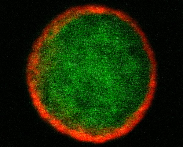
A chimeric antigen receptor (CAR)-T cell (in green) with the CAR molecule stained in red. Image: Paula Martí Torrell
Oncolytic viruses
Oncolytic viruses can induce immunogenic cancer cell death releasing endogenous alarm signals for activation of the immune system triggering anti-tumour T cell responses. Such viruses may therefore pave the way for effective immune checkpoint blockade and other immunotherapies.
We are developing oncolytic viruses armed with factors for recruitment and activation of dendritic cells that upon activation can take up tumour antigen from killed tumor cells and cross-present antigenic epitopes to prime cytolytic T cells.
Neuroendocrine Cancer
A recombinant oncolytic adenovirus, called AdVince, was developed in the lab. The virus capsid was modified for increased uptake in tumour cells and neuroendocrine virus replication is ensured by introduction of a specific promoter and microRNA target sequences in the virus genome. AdVince is currently being evaluated in neuroendocrine cancer patients at Uppsala University Hospital.
Glioblastoma
Semliki Forest virus (SFV) is a neurotropic virus that is being evaluated as an oncolytic agent in preclinical glioblastoma models. We restrict virus activity by insertion of microRNA target sequences in the virus genome and arm the virus with transgenes able to induce antitumoral immunity. We recently identified VLDLR as a crucial receptor for SFV entering the central nervous system. Other neurotropic viruses are also being evaluated.
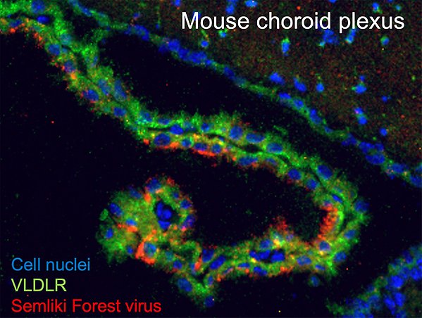
Semliki Forest virus (SFV) entering the central nervous system through infection of very low-density lipoprotein receptor (VLDLR)-positive cells in the choroid plexus of a brain ventricle. Cell nuclei are shown in blue, VLDLR is stained in green and the SFV is stained in red. Image: Miika Martikainen.
Viral vectors to improve cancer immunotherapy
We are, together with Anna Dimberg and her research group at Uppsala University, developing tumour vessel-targeted adeno-associated viral (AAV) vectors encoding factors able to transform tumour-associated endothelial cells into high endothelial venules (HEVs). The aim is to improve T cell recruitment into tumours and induction of antigen-presenting niches in the tumour microenvironment to sustain T cell activity.
The Dimberg lab, Thomas Tüting’s lab in Magdeburg University, and our lab are also trying to map and connect the vascular tumour landscape with the tumour immunity landscape.
We are now entering the phase of scaling up capsid modified AAV vector production to bridge the cap to clinical trials.
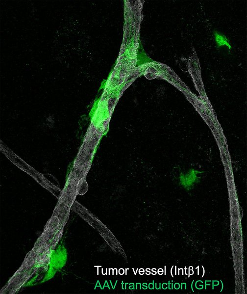
A specifically designed AAV vector carrying GFP transducing activated integrins on blood vessels in brain tumor. Vessels are indicated in gray and AAV transduction is shown in green. Image: Bertin Paranjothi Mary